By Madison Fratzke, RRT, RRT-ACCS, and Keith Lamb, RRT, RRT-ACCS, FAARC, FCCM
Introduction
Influenza can lead to respiratory failure which can evolve into significant Acute Respiratory Distress Syndrome (ARDS). Standard treatment for significant ARDS is invasive positive pressure ventilation which in and of itself may cause deleterious secondary inflammatory processes leading to a precipitous increase in mortality. It has been demonstrated that the implementation of Extracorporeal Life Support (ECLS) may mitigate these effects and improve outcomes in patients with severe respiratory failure caused by the influenza virus. 1
Influenza in the US
In the U.S. last year, influenza caused an estimated 79,000 deaths. While mortality varies each year, knowledge of infection prevention is well known and includes receiving an annual influenza vaccine. Still, the effectiveness of the vaccine is influenced by the pandemic nature of the virus, timing of its arrival to the U.S., and compliance with receiving the vaccine. Of the many complications that can occur in someone diagnosed with influenza, infection can lead to ARDS, another life-threatening condition. 1-2
Influenza-related ARDS
ARDS is predominantly an inflammatory process that can be caused by a primary or secondary trigger. Influenza acts as a primary etiology and when there is an exaggerated inflammatory response the result can be severe respiratory failure. As previously described, Influenza is a viral infection and as such does not respond to antibiotic therapy, but there are several approved antiviral medications prescribed in the U.S. These medications along with fluid management are the standard of care most often followed. In the face of severe respiratory failure, these are often not enough, and the use of invasive mechanical ventilation is needed to maintain adequate life-sustaining gas exchange. 3-4<s/up>
Mechanical Ventilation in ARDS
Since the year 2000, evidence has repeatedly shown that optimizing lung protective ventilation reduces stress and strain on the lungs, therefore reducing days on the ventilator and improving mortality. The utilization of lower tidal volumes, higher positive-end expiratory pressure and lower inspired oxygen levels has been shown to prevent secondary inflammatory processes by reducing the traumatic opening and closing of the distal airways and alveoli. The ARDSnet trial resulted in an ARDSnet protocol to assist in properly managing significant ARDS patients, but when injurious mechanical ventilation settings can no longer be avoided, the addition of other evidence-based treatment adjuncts should be considered without delay. 5-6
Indications for ECLS
As discussed above, there are risks associated with mechanical ventilation. At some point, and it is not completely understood when this occurs, these risks outweigh the benefit that can be afforded by the ventilator. This most likely occurs when there is excessive stress, strain and inspired oxygen levels delivered to the lungs. When respiratory failure is so severe that these injurious levels of mechanical ventilatory support are required to maintain adequate gas exchange, ECLS may be indicated. ECLS takes the emphasis off the mechanical ventilator and provides gas exchange so that harmful levels of support from the ventilator can be reduced thereby mitigating the injurious effects. The type of ECLS that is deployed to support respiratory function is Veno-venous Extracorporeal Membrane Oxygenation or ECMO. 7
Role of the Respiratory Therapist
Respiratory Therapists play a major role in the care of any critically ill patient but even more so when there is significant respiratory failure. RTs may serve as ECLS Specialist’s taking primary responsibility for the ECLS management and or ventilator management. Either way, it is imperative that protective strategies be implemented and adhered to by both the bedside RT and the ECLS Specialist.
Case Report
History of present illness:
A 21-year-old male who was feeling tired, achy with some shortness of breath presents to a local freestanding ER. His room air SpO2 is 75%, he has scattered crackles, is afebrile and normotensive, and is slightly tachypneic with a resting respiratory rate of 24.
He has no significant medical or surgical history.
Initial blood gas analysis reveals hypoxemia with mild respiratory alkalemia.
Labs revealed influenza A and B via filmarray.
Progression
Over the next two hours, the patient became more hypoxemic, tachypneic and distressed. He was placed on heated high flow nasal cannula, then on non-invasive ventilation and finally was intubated and placed on protective mechanical ventilator settings. Initial settings were Volume Assist Control, tidal volume 6 ml/kg/pbw, frequency 20 bpm, FiO2 100% and 10 cm H2O of PEEP.
Over the next few hours, the patient’s hypoxemia became worse and there was evolving respiratory acidemia. His plateau pressure with 6 ml/kp/pbw was 35 cm H2O. Tidal volumes were dropped to 4 ml/kg/pbw but plateau pressures continued to climb while the acidemia and hypoxemia also worsened.
The ICU team decided that in order to prevent the potential deleterious impact that such high ventilator settings may have on the patients’ outcome, ECMO would be a reasonable approach.
The patient was cannulated with a 31 French dual lumen, bi-caval cannula placed in the right internal jugular vein and veno-venous extracorporeal membrane oxygenation was initiated.
Ventilator settings were reduced immediately, and his compliance improved over the next few days allowing further reduction of support.
The patient remained on VV ECMO for 6 days.
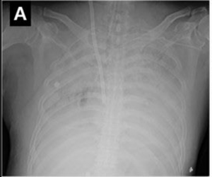
Compliance, gas exchange, and the chest film all improved, and the patient was decannulated on day 7, extubated on day 8, and discharged home on hospital day 11.
Clinic follow-up revealed a normal baseline 21-year-old male.
Summary
Influenza respiratory infections can rapidly evolve into life-threatening respiratory failure. When primary and secondary inflammatory processes occur in patients with influenza-related ARDS, ECLS should be considered to allow the patient time to recover while preventing further lung injury. 1-7
- Jaber, S., et al. “ARDS and influenza A (H1N1): patients’ characteristics and management in intensive care unit. A literature review.” Annales françaises d’anesthèsie et de rèanimation. Vol. 29. No. 2. 2010.
- Center for Disease Control. (2018) Influenza. Retrieved from https://cdc.gov/flu/about/index.html
- Grasso, Salvatore, et al. “ECMO criteria for influenza A (H1N1)-associated ARDS: role of transpulmonary pressure.” Intensive care medicine 38.3 (2012): 395-403.
- Pappalardo, Federico, et al. “Predicting mortality risk in patients undergoing venovenous ECMO for ARDS due to influenza A (H1N1) pneumonia: the ECMOnet score.” Intensive care medicine 39.2 (2013): 275-281.
- Acute Respiratory Distress Syndrome Network. “Ventilation with Lower Tidal Volumes as Compared with Traditional Tidal Volumes for Acute Lung Injury and the Acute Respiratory Distress Syndrome” New England Journal of Medicine. 2000.
- Spieth, P. M., A. Güldner, and M. de Abreu Gama. “Acute respiratory distress syndrome: Basic principles and treatment.” Der Anaesthesist 66.7 (2017): 539-552.
- Bartlett, Robert H. “Extracorporeal membrane oxygenation for acute respiratory distress syndrome: EOLIA and beyond.” Critical care medicine 47.1 (2019): 114-117.
Email newsroom@aarc.org with questions or comments, we’d love to hear from you.
Heather Willden is the Director of Governance and Strategic Initiatives for the AARC where she works with state affiliates as the HOD liaison. She also manages DEI efforts and strategic initiatives. Connect with her about these topics by email, AARConnect or LinkedIn. When she's not working, you can find her podcasting with her husband, exploring new hiking trails, photographing, and spending time with her family.